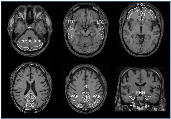
Fig. (1)
Region of Interest (ROI). Each ROI was manually delineated on standard MR images to which individual PET data was standardized after acquisition of individual MR images. LTC: Lateral Temporal Cortex, FRC: Frontal Cortex, PCG: Posterior Cingulate Gyrus, PC: Precuneus, PAR: Parietal Cortex, PHG: Parahippocampal Gyrus.