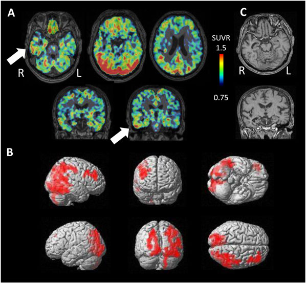
Fig. (6)
A case with AGD. (A) Tau accumulation was found on the right temporal lobe (arrows) and parietal lobe. (B) Cortical projection reveals significant accumulation of tau diffusely on the right parietal lobe. (C) MRI reveals hippocampal atrophy on the right side.