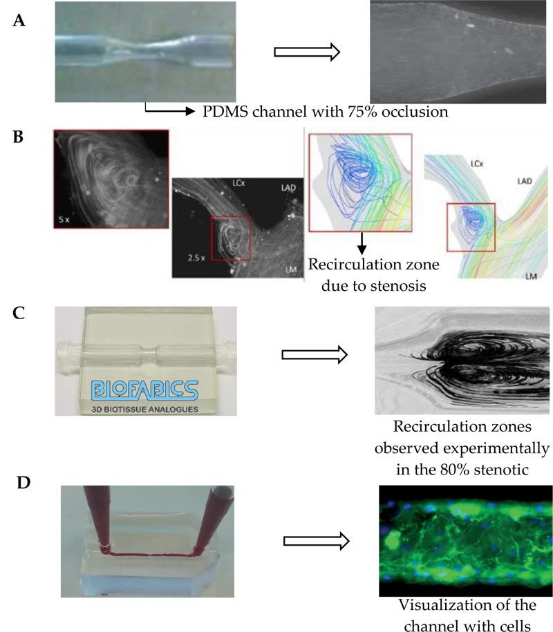
Fig. (4)
Examples of some biomodels and respective experimental images: A) Rigid PDMS phantom and flow visualization of a stenotic artery, adapted from [83]; B) Flow visualization and streamlines obtained numerically of a stenotic left coronary artery, adapted from [75]; C) Stenotic model fabricated by Biofabics and the recirculation zones observed experimentally, adapted from [63]; D) PDMS model of a stenotic coronary artery and image obtained by means of fluorescence microscopy, adapted from [77].