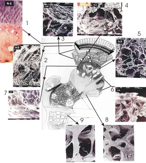
Fig. (1) shows a schematic drawing of a bone with its most characteristic fine structure (shown as SEM pictures with micrometer resolution): 1: cartilage, 2: tide –marrow, 3: calcified zone, 4: subchondriala compacta, surface of femur head, 5: cysterns between calcified zone, 6: subchondriala compacta (inverted), 7: epi-line (epiphysal joint): 8: Paul`s pressure-tension trajectories, 9: transition between joint and diaphysis.