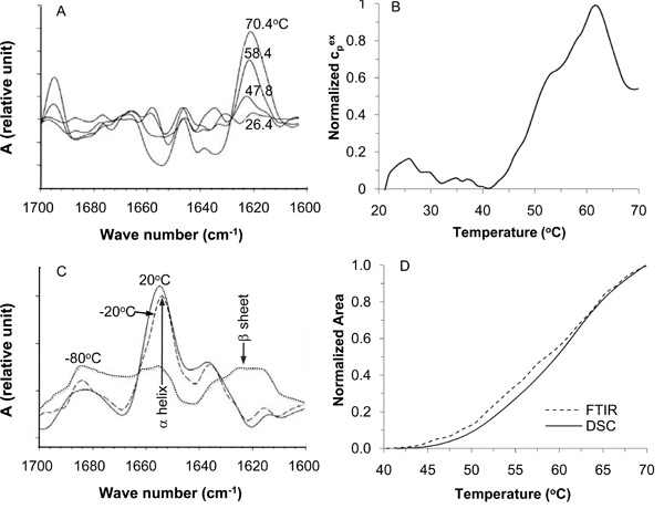
Fig. (6) Fourier transform infrared (FTIR) and differential scanning calorimetry (DSC) signatures of thermally induced changes in cellular proteins in situ: (A), FTIR spectra at different temperatures showing the increase of extended β sheet structure and a simultaneous decrease of the α helix structure in cellular protein during heating mammalian cells slowly (2 °C/min); (B), DSC endotherm demonstrating heat absorption as a result of protein denaturation during heating mammalian cells slowly also at 2 °C/min; (C), FTIR spectra showing extensive change of protein secondary structure from α helix to extended β sheet in thawed cells after freezing slowly to -80 °C, but not -20 °C; and (D), the DSC and FTIR measurements match well, suggesting protein denaturation is one of the major events that result in cell injury during heating between 40-70 °C. Figure reprinted and redrawn from references [158,195] with permission from Elsevier (for panels A and C) and Springer (for panels B and D).