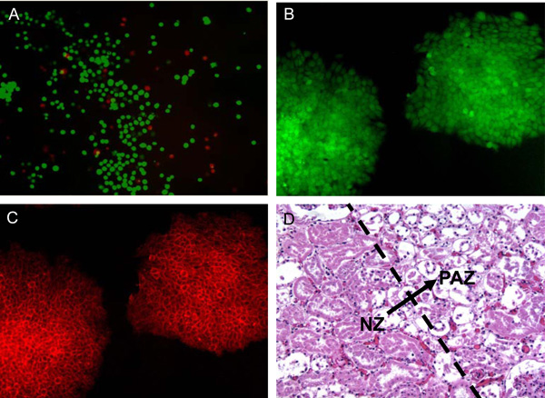
Fig. (7) Viability and function of mammalian cells post thermal treatment evaluated by exemplary assays: (A), immediate viability of cells assayed using calcein AM (green fluorescence indicating cellular metabolic activity and intact membrane of viable cells) and ethidium homodimer (red fluorescence indicating compromised cell membrane of injured and potentially dead cells); (B) and (C), expression of green fluorescent protein (GFP) under the control of Oct-4 gene (B) and a membrane surface glycoprotein (SSEA-1, C) indicating the undifferentiated properties of R1 murine embryonic stem cells post thermal treatment; and (D) histological difference between viable and damaged renal cells in normal porcine kidney tissue after 2 day’s culture in media post thermal treatment showing the transition (arrow) from the lower left zone of significant necrotic tissue to the upper right zone with mainly intact tissue: Damaged cells lack nuclei. Figure reprinted and redrawn from references [38,64] with permission from Elsevier (for panels B and C) and Informa (for panel D).