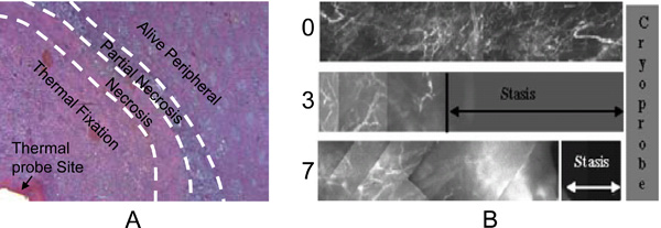
Fig. (8) Both histology and blood perfusion defects have been used to evaluate tissue injury in vivo: (A), a micrograph showing the patterns of in vivo hyperthermic injury in porcine renal tissue 7 days after thermal treatment and (B) in vivo blood perfusion defects due to cryothermic injury in prostate tissue at different times (i.e. 0, 3 and 7 days) measured in situ by fluorescence contrast. Although wound healing was observed in the necrotic and partially ablated zone, a central lesion named thermal fixation knowing for its resistance to wound healing response were observed in kidney tissue exposed to hyperthermic temperatures (A). Wound healing was observed throughout the cryogenic lesion leading to its shrinkage from day 3 to day 7 (B). Figure reprinted and redrawn from references [38,247] with permission from Informa (for panel A) and ASME (for panel B).