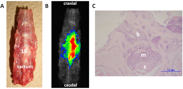
Fig. (3) Ex vivo imaging and histological examination of the dissected spine (Rat No. 7). The dissected lumbar spine was set in the supine
position (A). Fluorescence can be clearly detected in the metastatic lesion in the L6 vertebra (B). Histological examination (C) of the axial
cross-sections of the L6 vertebra (H&E staining, scale bar = 1 mm) shows invasion of the vertebral bone marrow spaces by cancer cells (b).
The metastatic lesion destroyed the trabecular bone structures of the spinal canal (m), and compressed the spinal cord (c) on day 11 after
tumor implantation.