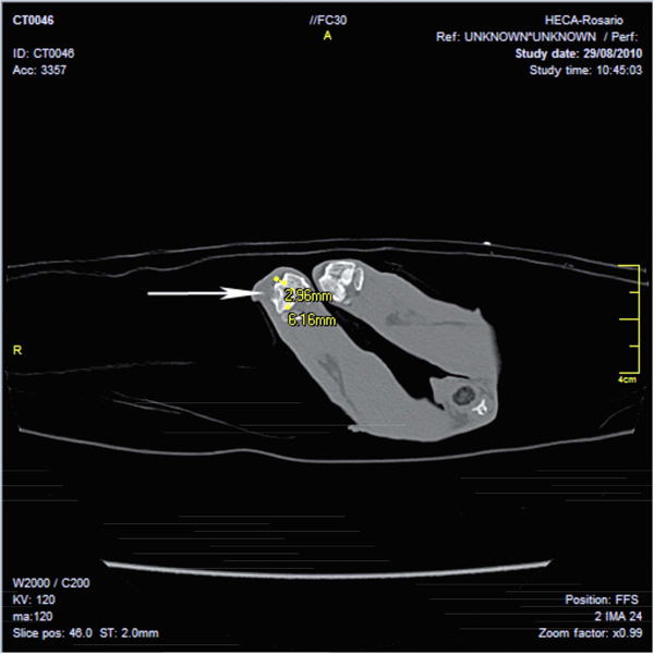
Fig. (5) Image obtained in Multislice CT scanner Toshiba (16 slice) shows a bone lesion in a rabbit femur according to material and method.
A radiolucent image with cortical erosion in distal femoral epiphysis of right condyle is observed. Dense image interpreted as signs of
incipient integration-regeneration. No swelling of soft tissue is seen.