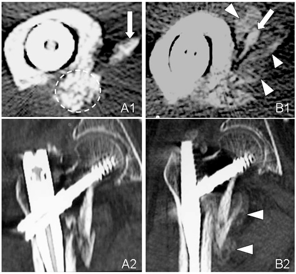
Fig. (4) Axial (A1, B1) and sagital (A2, B2) CT images of the unstable intertrochanteric fracture in Case 1. The complex of β-TCP granules,
hyaluronate, and rhFGF-2 remains in its original place (broken circle) at 3 weeks (A1). CT image shows marked new bone formation at 6
weeks (B1, B2). The arrows point to the lesser trochanter and arrow heads point callus.