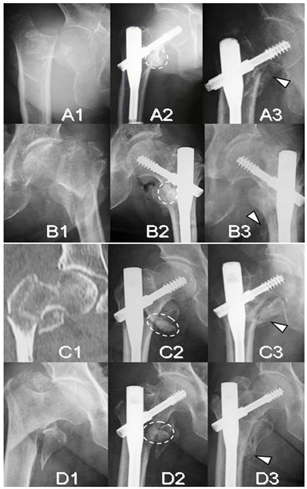
Fig. (5) Radiographs and a CT image of four unstable intertrochanteric fracture cases (A;Case 2, B;Case 3, C;Case 4, and D;Case 5). Initial
radiographs (A1, B1, C1, and D1). Immediate postoperative radiographs (A2, B2, C2, and D2). Follow-up radiographs at 12 weeks (A3, B3,
C3, and D3). Broken ovals indicate the remaining complex of β-TCP granules, hyaluronate, and FGF-2 and arrow heads indicate callus.