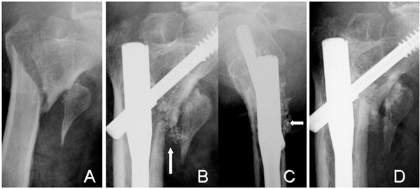
Fig. (6) Radiographs of an 83-year-old woman (Case 6) with a displaced lesser trochanter (A). The β-TCP granules are still visible at 4
weeks (arrows) in AP (B) and lateral views (C). The complex is resorbed and bone union of the intertrochanteric fracture has occurred at 12
weeks (D). However, no bridging bone formation is seen between the lesser trochanter and the shaft.