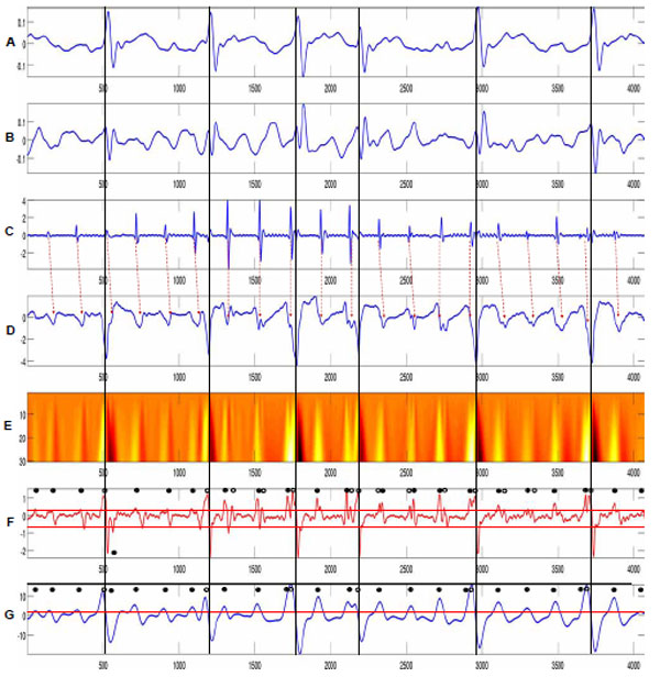
Fig. (3) The proposed wavelet analysis method was applied to the atrial electrograms at the site I in the LA anterior wall of a patient with PAF. (A) ECG I lead. (B) ECG aVF. Starting point of QRS event is denoted by dotted vertical lines. Atrial electrograms were collected by distal of ablation catheter (bipolar) and non-contact EnSite array (unipolar) simultaneously at the site I in (C) and (D), respectively. The scalogram for the unipolar electrogram using wavelet transforms was displayed in (E). Weighted fine and coarse scale electrograms ffine and fcoarse were constructed in (F) and (G), respectively.

