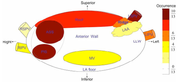
Fig. (7) Schematic representation of occurrence in the thirteen patients of upper quartile frequencies by region in the LA (the anterior view). The colour spectrum displays occurrence of electrical activation. LAA = left atrial appendage; RLA = right lateral wall; LLW = left lateral wall; ASS = antero-superior septum; PIS = postero-inferior septum; LSPV = left superior pulmonary vein; LIPV = left inferior pulmonary vein; RSPV = right superior pulmonary vein; RIPV = right inferior pulmonary vein; MV = mitral valve. The ridge is the triangle region between the LA roof, LSPV and LAA. PLA = posterior left atrium.