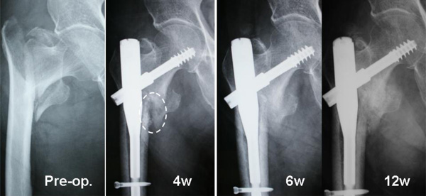
Fig. (3) Radiographs of an
82-year-old woman (Case 1) with a displaced lesser trochanter. The injected
complex remained in its original place (white oval) at 4 weeks. Callus formation
was found 4 weeks after surgery and the callus increased in size over time.
Marked new bone formation was found around the lesser trochanter at 6 andRadiographs
of an 83-year-old woman (Case 6) with a displaced lesser trochanter (A).
The β-TCP granules are still visible at 4 weeks (arrows) in AP (B) and
lateral views (C). The complex is resorbed and bone union of the
intertrochanteric fracture has occurred at 12 weeks (D). However, no
bridging bone formation is seen between the lesser trochanter and the shaft. 12
weeks.