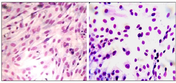
Fig. (9) Micrographs of fibroblastic cells on the surface after 5 days cultivation during in vitro tests. Left: double-layered alumina/calcium-phosphate coating on a Ti6Al4V substrate. Right: area with separated cells and naked-nuclear cells on an uncoated Ti6Al4V substrate. Staining with hematoxylin and eosin, magnification ×400.