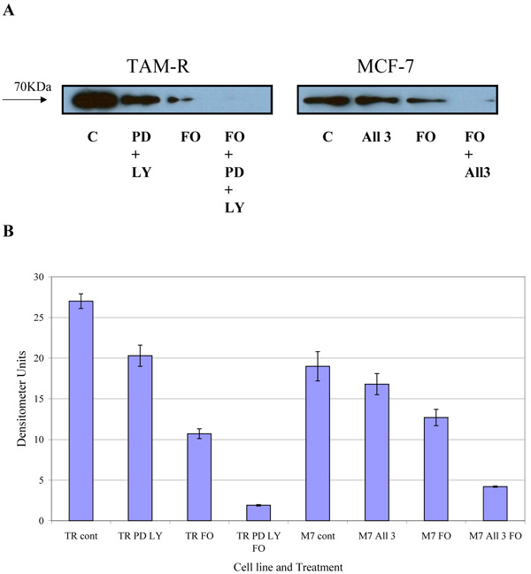
Fig. (6) Western blot analysis of the levels of total COX-2. MCF-7 and TamR cells were incubated with FO (1 μM L-1 fish oil), PD (25 µM PD98059), LY (5 µM LY294002) and 1 x 10⁻⁷ M 4-hydroxytamoxifen (MCF-7 cell only) for 3h. Cells were lysed and protein extracted and quantified for equal loading. Protein was separated by SDS-PAGE. Proteins were immobilised on a nitrocellulose membrane and immunoprobed using a total COX-2 primary antibody. B. Densitometric analysis of total COX-2. Densitometry was performed using Alpha Digi Doc TM RT camera and image analysis system, Genetic Research Instrumentation, Essex, UK. M7- MCF-7 cells, TR- TamR cells (n=3 ± SD). Passage numbers were 23, 27 and 30.