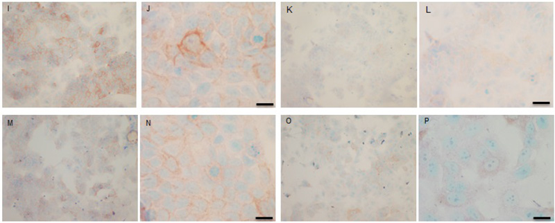
Fig. (9 I-P)
Immunocytochemical detection of total COX-2 in TAMR cells. Cells were incubated with 0.1 µL mL-1 DMSO (I & J), 1 µL mL-1 fish oil (K & L), 25 µM PD98059 and 5 µM LY294002 (M & N) or 1 µL mL-1 fish oil, 25 µM PD98059 and 5 µM LY294002 (O & P) for 3h. Cells were grown to 70% confluency on TESPA coated glass coverslips and fixed using formal saline. Primary antibody was NEB total COX-2 rabbit primary antibody at 1/40 for 1.5h. Secondary antibody was DAKO Rabbit EnVision peroxidase labeled polymer antibody for 1.5h. Magnification: I, K, M, O x 10, L x 20 and J, N, P x 40, scale bar = 50 μm. Passage numbers were 15, 19 and 30.