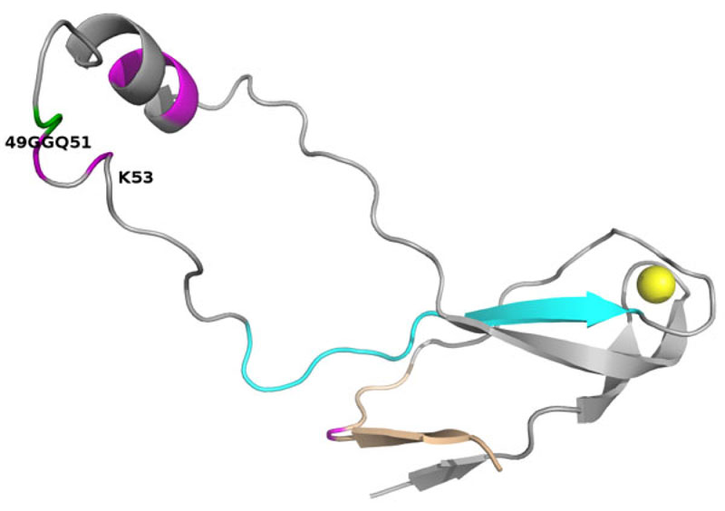
Fig. (5)
Model of the human eL42 protein. Ribbon representation of the 3-D structure of human rp eL42 (formerly RPL36AL) (fragment 1-94) modeled by homology with the crystallographic structure of the archaeal counterpart rp eL42 (formerly RPL44E) of the 50S ribosomal subunit from Haloarcula marismortui [28, 29]. The post-translational modifications (including the methylated Q51), and the consensus pattern 61Kx(TorV)KKxxL(KorR)xxC72 (numbering of human eL42) of the eL42 family of r-proteins are colored pink and cyan, respectively. The 49GG50 dipeptide of the GGQ motif is highlighted in green. A zinc ion represented by a cadmium colored yellow is also shown. Fragment 86-94 corresponding to the nucleotide binding motif 2 (NBD2) is shown in wheat.