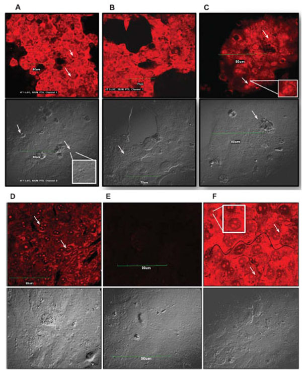
Fig. (2). Paclitaxel induces nuclear fragmentation and cell death in 4T1-Luc tumor cell lines The extent of DNA damage observed in tumor cells was dose dependent. Arrows point to fragmented nuclei. Inserts highlight nuclear fragments and damaged DNA within cells. Scale bar indicates 90 µm. Cell morbidity was determined by Live-Dead Viability Assay. Drug treated cells were incubated with ethidium homodimer (EthD-1), which stains fragmented nuclei of dead cells. EthD-1 dye can only penetrate cells with compromised membranes and emits red fluorescence (ex/em - 495 nm/ 635 nm) on binding to nucleic acids. Exclusion of the dye by intact membrane of live cells distinguishes dead cells from live ones. (A – F) - Apoptosis in 4T1-Luc cells. Cells treated with high paclitaxel doses, 50 µM (A) and 25 µM (B), show extensive nuclear fragmentation and damaged membranes. (C) At 12.5 µM and (D) 6.25 µM, moderate nuclear fragmentation is observed. (E) Reflects background fluorescence from negative control cells. (F) Apoptotic induction in positive control (ethanol-treated) cells. Wavy black line traces boundary of damaged cell membrane.