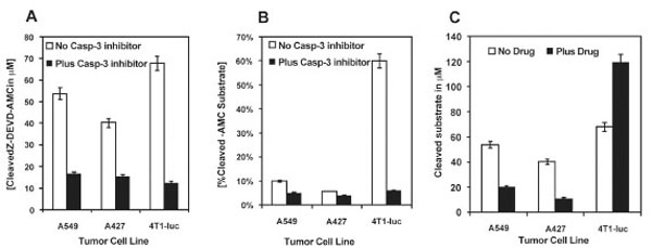
Fig. (4). Tumor cell lines exhibit differential basal and drug-induced caspase-3 activity The intrinsic and drug-induced caspase-3 activity of 3-cell lines were determined by a fluorimetric enzymatic assay that measures cleavage of a caspase-3 specific substrate, Z-DEVDAMC,where Z represents a benzyloxycarbonyl group, and AMC, 7-amino-4-methylcoumarin. The substrate fluoresces weakly in the UV range (Ex/Em~330/390 nm) but yields strong blue-fluorescent product (Ex/Em ~ 342/441 nm) upon proteolytic cleavage and allows continuous monitoring of the activity of caspase-3 and its related proteases in cell extracts. A standard AMC-substrate curve was used to quantify the levels of enzyme-cleaved substrate from the measured fluorescence values. To confirm that the observed fluorescence signal was due to the activity of caspase-3-like proteases, control treatments were incubated with 10 M of a reversible caspase-3 inhibitor, Ac-DEVD, prior to treating cells with the substrate. Experiments were performed twice in triplicate. N= 6. Data show mean ± SEM of 2 experiments done in triplicate.(A) While all 3-lines showed some level of basal enzymatic activity, A427-cells showed the least activity with 40 µM of cleaved substrate, followed by A549-cells, with 54 µM of cleaved substrate. 4 T1-Luc cells showed the highest levels of activity with 68 µM of cleaved sub-strate. A decrease in activity in the presence of a caspase-3 inhibitor confirms the specificity of the reaction. The underlying difference in caspase-3 activity may account for the observed resistance patterns after drug exposure. (B) Paclitaxel induces differential caspase-3 activity in tumor cell lines. Whereas 4T1-Luc cells showed an increase in levels of cleaved product (120 µM, 60% of added substrate) after exposure to drug, A549 and A427-cells showed a decrease in the levels of cleaved product, with 20 µM and 11 µM, respectively, of cleaved substrate. This suggests that while 4T1-luc cells activate caspase-3 on exposure to drug, A549 and A427 cells somehow repress caspase-3 activation. (C) In comparison to basal enzymatic activity levels, paclitaxel induces a two-fold increase in caspase-3 activity in 4T1-luc cells as measured by increased levels of cleaved substrate. In contrast, A549 and A427 show approximately a 3-fold (2.7) and 4-fold (3.64) decrease in enzy-matic activity, respectively. This increased activation of caspase-3 underlies the sensitivity of 4T-1uc cells to drug, whereas, repressed cas-pase-3 activation potentially accounts for the resistance of A549 and A427 cells to paclitaxel