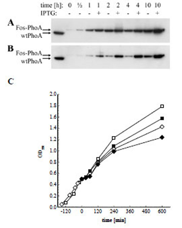
Fig. (3) Western blot analysis of periplasmic extracts directly before, 1/2 h, 1 h, 2 h, 4 h and 10 h after helper phage superinfection, and of soluble wtPhoA (left lane). (A) Fos-PhoA detected in XL1-Blue harbouring pJuFoIII::phoA without (-) and in the presence of 1 mM IPTG (+). (B) Fos-PhoA detected in XL1-Blue harbouring pJuFoVIII::phoA grown without (-) and in the presence of 1 mM IPTG (+). (C) Growth rate of XL1-blue/pJuFoIII::phoA (squares) and XL1-blue/pJuFoVIII::phoA (circles) grown without (open symbols) and with 1 mM IPTG (solid symbols)