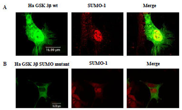
Fig. (3). Confocal microscopic analysis of GSK 3β wt or its SUMO mutant Confocal microscopic analysis of transfected Ha –GSK 3β wt (A), Ha –GSK 3β SUMO mutant (K292R) (B) was performed to determine whether it merged with SUMO-1 (red color). All Ha –GSK 3β constructs were shown as green color. The transfected Ha -GSK 3β wt (de-tected in both the cytoplasm and the nucleus) merged (yellow) with SUMO-1 in the nucleus (A). The transfected Ha –GSK 3β SUMO mutant was detected in the cytoplasm, but not in the nucleus (B). The SUMO-1 modification proteins were mainly detected in the nuclear region (B middle lane). GSK 3β SUMO mutant in which the SUMOylation site was eliminated was not merged with SUMO-1 in the nucleus (B right lane).