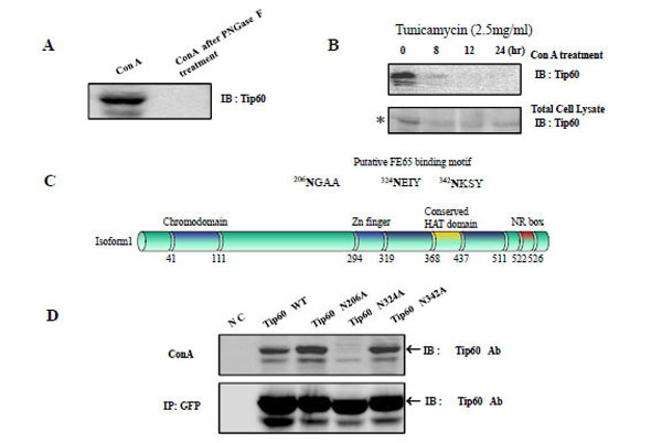
Fig. (1) The association Tip60 with concanavalin A in Vivo and the putative Site of Tip60 N-glycosylation A) After treatment HEK293 cell lysate with concanavalin A (Con A, Jack bean lectin), the extracts were then analyzed by Western blotting using an anti -Tip60 antibody (A left). To control the N-glycosylation of Tip60, we also added Con A to HEK293 cell lysate was pretreated with PNGase F, before Con A treatment (A right). B) The reactivity of Tip60 to Con A was reduced with the tunicamycin (2.5mg/ml) treatment for different times, as indicated above (B upper lane). To control the amount of Tip60, the each cell lysate was immuno blotted with its specific antibody (B bottom lane). * indicated the putative glucosylated Tip60. C) With the http://www.cbs.dtu.dk/services/SignalP/output.html program, the putative N-glucosylation sites in Tip60 was predicted, as indicated in Fig. 1C above. The domains (chromodomain for the histone binding, Zn finger, the conserved HAT domain, NR for the nuclear receptor binding) are also indicated [6, 7]. D) After transfected with GFP Tip60 WT, N206A, N324A, N342A, the cell lysates was precipitated either Con A (D upper lane) or GFP Ab (D bottom lane). To detect Tip60 amount, immunoblot was performed with its specific Ab.