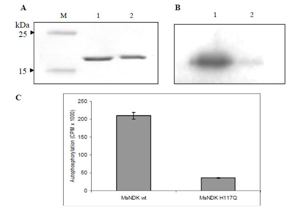
Fig. (6) Autophosphorylation assay of MsmNDK and MsmNDK-H117Q. A. Coommassie blue stained SDS-PAGE gel profile. Lane M, Mol
Wt markers. Lane 1, MsmNDK (1 µg). Lane 2, MsmNDK-H117Q (1 µg). B. Autoradiograph of MsmNDK (lane 1) and MsmNDK-H117Q
(lane 2) after autophosphorylation. C. Bar graph represents the quantitation of autophosphorylation of MsmNDK and MsmNDK-H117Q.
Assay was repeated independently three times for each sample. Counts per minute (CPM) were recorded and standard deviations were calculated.