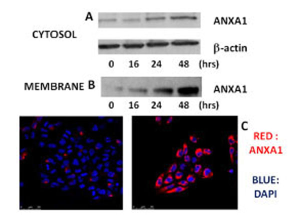
Fig. (3) Effects of xanthohumol on the expression of ANXA1 in the cytosol and on the membrane of human T98G glioblastoma multiforme cells. (A) Analysis of the expression of ANXA1 in the cytosol of T98G cells. (B) Expression of levels of ANXA1 on the plasma membrane of cells T98G. T98G cells were incubated in the presence or absence of xanthohumol (20µM) at different times. The expression of ANXA1 was analyzed by Western blotting using a polyclonal anti-ANXA1. (C) Expression of levels of ANXA1 on the plasma membrane of T98G cells analyzed by confocal fluorescence microscopy. After 24 hours of incubation the cells were treated with 20µM xanthohumol for the stated time, subsequently the cells were washed and incubated with a polyclonal antibody anti-ANXA1 and with DAPI to have a colored of the nuclei. The intense red color that is seen in the figure is indicative of the presence of ANXA1 on the membrane.