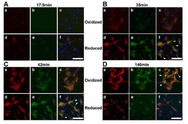
Fig. (2) Redox conditions affect reductide uptake and intracellular fluorescence.Live cell time-lapse
confocal microscopic images (60X) of BJ fibroblasts incubated with reductide are shown. Cells were
seeded into 4-chamber glass cover slides and allowed to attach overnight. The following day, the plates
were pretreated with either CDNB 25 µM or NAC 4 mM for 30 minutes prior to washing with PBS and
incubating with reductide 4 µM in normal media. TAMRA emission images are shown in red, FAM images
are shown in green, and DAPI images are shown in blue. Arrows indicate exocytic vesicles containing
FAM but not TAMRA.