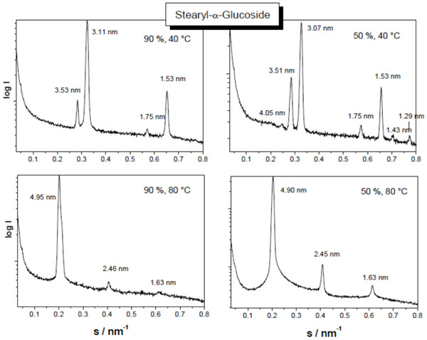
Fig. (19) Small-angle X-ray scattering patterns of stearyl glucoside at 90 % (left spectra) and 40 % (right spectra) water content, and at 40 °C
(top spectra) and 80 °C (bottom spectra). The logarithm of the scattering intensity is plotted versus the scattering vector s (=1/d, d = spacings
of the reflections). From Vill et al., Chem. Phys. Lipids 104, 75-91 (2000), with permission by Elsevier.