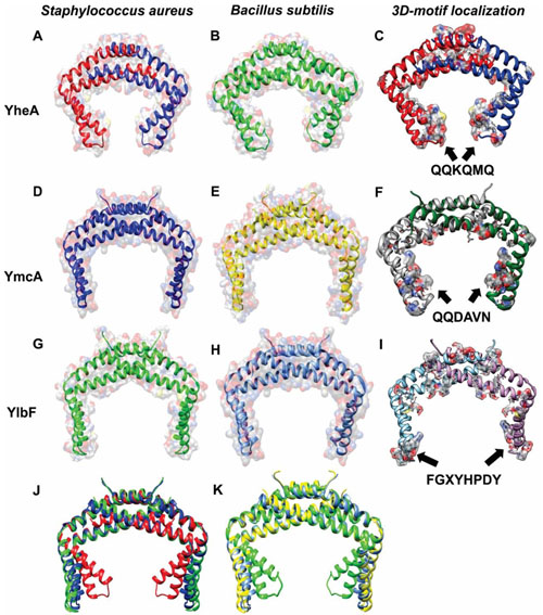
Fig. (3) Tridimensional structure models of the YheA, YmcA and YlbF proteins identified in Staphylococus aureus and Bacillus subtilis and localization of their putative motifs.Homodimer structures of the three proteins showing its solvent accessibility and location of their putative motives. A. YheA protein, B. YheA protein modeling from partial tertiary structure reported in the PDB database (2OEE), C. Staphylococcus aureus YheA protein with its putative motif QQKQMQ (Black arrows), D. YmcA protein, E. YmcA protein reported in the PDB database (2PIH), F.Bacillus subtilis YmcA protein with its putative motif QQDAVN, G y H. Partial YlbF proteins, I.Staphylococcus aureus partial YlbF protein with its putative motif FGXYHPDY, J and K. YheA, YmcA and YlbF 3D models superposition.