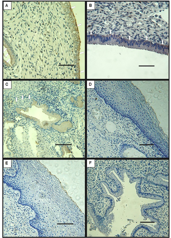
Fig. (1) Representative microphotographs of immunohistochemical analysis for DMBT1 of normal cervix biopsies. A, B- endocervix, arrows show specific staining at supranuclear (A) and infranuclear (B) regions. C- gland positive for DMBT1, arrows show metaplastic cells negative for DMBT1 staining. D- exocervix, E- transition zone, both negative for DMBT1. F- control using pre-immune serum. Bars indicate 50 μm (A, B) and 100 μm (C, D, E, F).