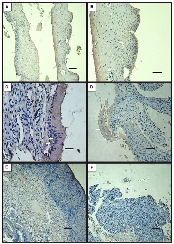
Fig. (2) Representative microphotographs of immunohistochemical analysis for DMBT1 of cervix biopsies with different grades of lesion. A- exocervix from LSIL biopsy, B- 2x amplification of A showing koilocytes. C- endocervix of columnar epithelium with infranuclear DMBT1 specific staining (arrows) in LSIL specimen. D- HSIL specimen showing recognizable detached endocervix monolayer epithelium, positive for DMBT1 staining (arrows). E- exocervix and glands in HSIL specimen. F- SCC with non-recognisable cervix regions. Bars indicate 100 μm (A, E, F), 50 μm (B, D) and 25 μm (C).