Fig. (1)
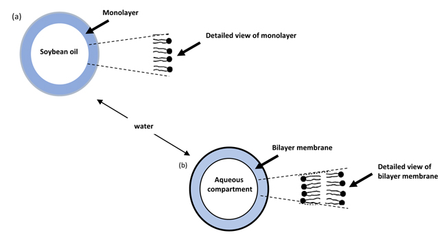
(a)
Schematic drawing of an Intralipid-10% particle after sonication.
(b)
Model of a lipid vesicle in the water environment.