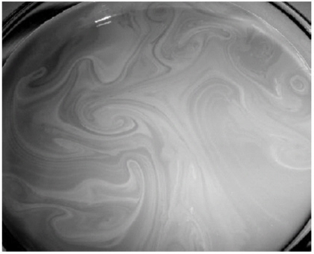
Fig. (3)
Surface layer on a water-Intralipid-ink dilution (μa ≈ 0.3 mm-1 and μs′ ≈ 3 mm-1) formed after 15 min of standstill and subsequent gentle stirring. This camera image is enhanced in contrast to improve the visibility of the flow marks. Reprinted with permission from Bodenschatz et al. [32]. © Institute of Physics and Engineering in Medicine. Reproduced by permission of IOP Publishing. All rights reserved.