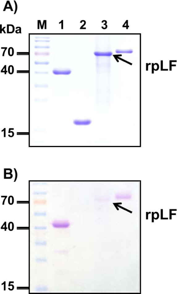
Fig. (4)
Glycosylation stain polyacrylamide gel electrophoresis.
A) Polyacrylamide gel stained with Coomassie blue; the same amount of horseradish peroxidase (lane 1, positive control), soybean trypsin inhibitor (lane 2, negative control), rpLF (lane 3, indicated by arrow) and bLF (lane 4) was subjected to SDS-PAGE and stained with Coomassie blue. Lane M, molecular mass markers.
B) The same amount of protein samples from A) stained with periodic acid-Schiff. Lane M, molecular mass markers.