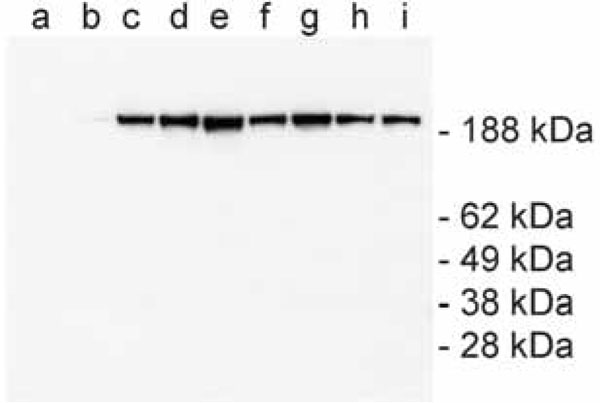
Fig. (2) Western blot analysis of dysferlin-TAP expression using a
complex of PAP. Equal amounts of total cell lysates were loaded in
each lane. Endogenous dysferlin is not recognized by PAP (lane a),
and only the recombinant dysferlin-TAP tag was detected (lane b-i).
Increasing amounts of the virus have been used for the transfection
of the cells in lane b to i: b: 10 µl, c: 25 µl, d: 50 µl, e: 100 µl, f: 200
µl, g: 300 µl, h: 400 µl, i: 500 µl.