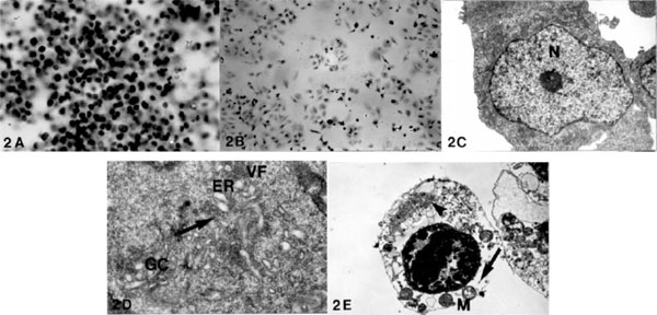
Fig. (2) Photomicrograph of NB cells in the control culture at 32 hours shows numerous cells with small and dark nuclei (A: Giemsa stain,
X 960). At the same time, the infected culture shows 60% CPE (B: Giemsa stain, X 960). Ultrastructure of the control culture at 20 hours
shows a large nucleus (N) containing a prominent nucleolus and a small amount of cytoplasm with preserved subcellular organelles (C: X
21,000). In the infected cells, the initial changes are observed in proliferation and dilation of ER and GC and VF at 20 hours. The vesicles are
originated from ER (arrow) (D: X 24,000). At 24 hours, the infected cultured cells show severe degeneration with a marked loss of the
ribosomes (arrow), VF (arrowhead) and alterations of the mitochondria (M) (E: X 22,000).