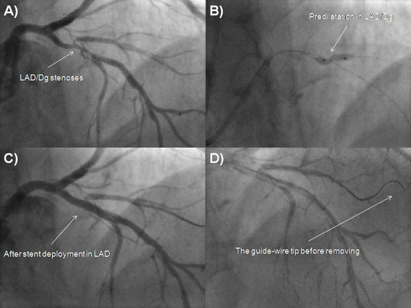
Fig. (1)
Fluoroscopic images of left coronary artery during PCI. A - Cine image of LAD/Dg stenosis. B - Cine image of LAD/Dg stenoses during predilatation. C - Cine angiography of LAD/Dg after PCI. D - The guide-wire tip before removal from Dg. Dg, diagonal branch; LAD, left anterior descending; PCI, percutaneous coronary intervention.