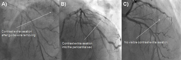
Fig. (2)
Fluoroscopic images of Dg perforation.
A
- Contrast extravasation projecting distal part of Dg.
B
- Contrast extravasation in the pericardial sac.
C
- The cessation of contrast extravasation in cine angiography. Dg, diagonal branch.