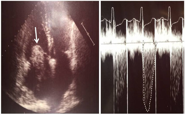
Fig. (2)
Two dimensional transthoracic echocardiography demonstrates a huge left atrial myxoma attached to the interatrial septum and prolapsing into the left ventricle during diastole. Mitral valvular stenosis and moderate mitral valvular insufficiency in a female patient (white arrow). Continuous doppler image of mitral valvular stenosis indicating a peak pressure difference of >67 mmHg between the left atrium and left ventricle.