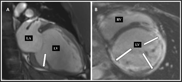
Fig. (3)
Cardiac MRI findings: 2-Chamber view (A) and short-axis view (B), showing a dilated left ventricle and wall thinning, especially of the inferior wall (A, arrow). Contrast-enhanced image (B) demonstrates extensive subepicardial late gadolinium enhancement, mainly in the inferior and lateral walls, and transmural uptake in the anterior wall (arrows). LA, left atrium; LV, Left ventricle; RV, Right ventricle.