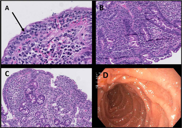
Fig. (4)
Histology of duodenal biopsy consistent with celiac disease, Marsh 3c, Hematoxylin-Eosin (H-E) stain: Illustrating intraepithelial lymphocytes (A, arrow), crypt hyperplasia (B), and severe villous atrophy (C). Gastroduodenoscopy (D) of the proximal duodenum showing normal macroscopic appearance except for slight oedema.