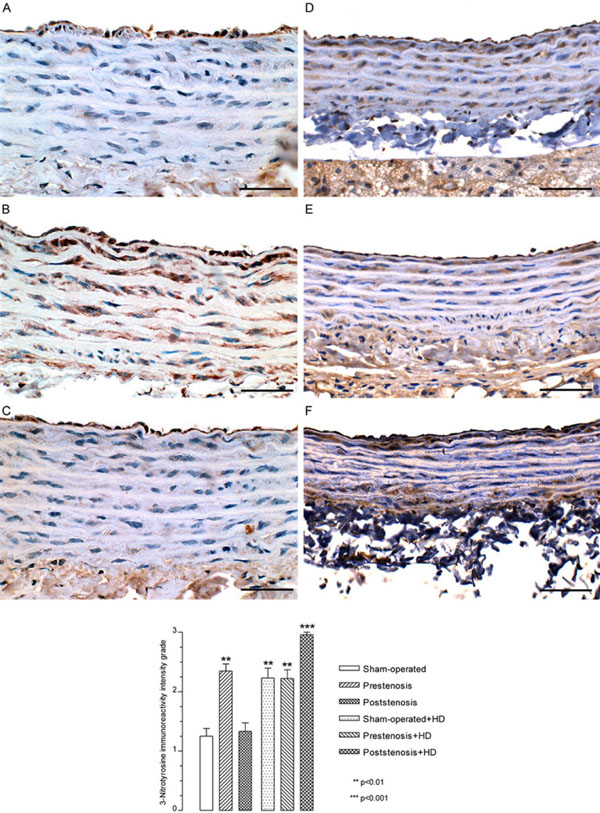
Fig. (5) Immunohistochemistry. Representative views of the aortas from sham-operated rats (A), prestenotic (B) and poststenotic (C) segments from operated rats and sham-operated+HD (D), prestenotic (E) and poststenotic (F) segments from operated+HD. The graph represents the quali-quantitative evaluation of the 3-nitrotyrosine immunoreactivity grade. The analysis revealed an increased expression of 3-nitrotyrosine in endothelial cells and smooth muscle cells in the prestenotic segment from operated group, sham-operated+HD, prestenosis+HD and poststenosis+HD as compared to sham-operated group. Scale bars, 50 µm.