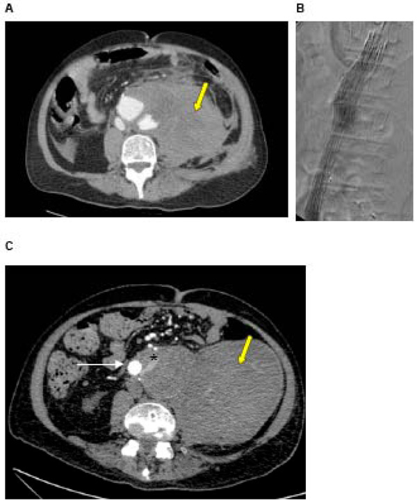
Fig. (2) A: CT of a ruptured AAA in a 72 year old man with a large haematoma (yellow arrow) B: Intra-operative angiogram during urgent EVAR showing stent graft in position, excluding the AAA C: Post-EVAR CT showing stent graft in situ and type II endoleak. Contrast is seen within the sac outside of the stent graft (white arrow = stent graft, * = endoleak), arising from one of the lumbar arteries. The haematoma caused at the time of rupture is still present (yellow arrow).