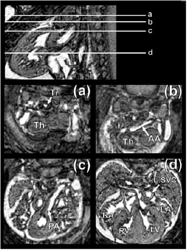
Fig. (12) Normal mouse embryo heart anatomy at 15.5 days post coitum (dpc) as seen by µMRI. a-d show successive axial slices through the thorax at the levels indicated. Key: Tr=Trachea, Th=Thalamus, AA=Ascending Aorta, PA=Pulmonary Artery, L.SVC=Left Superior Vena Cava, LA=Left Atrium, RA=Right Atrium, LV=Left Ventricle, RV=Right Ventricle. Note: dimension of heart is approximately 2 mm in diameter.