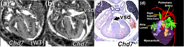
Fig. (13) Axial slices through Chd7+/+ and Chd7+/- embryos as part of a high throughput MRI screen. (a) Image showing a wild-type embryo with an intact ventricular septum. (b) image from an Chd7+/- embryo showing a ventricular septal defect (VSD) which was confirmed on later H&E histology (c) [134]. Volume rendering (d) of the same Chd7+/- heart and great vessels allows visualisation of the VSD (red triangle) in 3 dimensions.