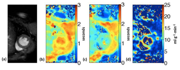
Fig. (4)
ASL perfusion imaging in the mouse. Short-axis image
(a)
, T
1
maps from global inversion
(b)
and slice-selective inversion
(c)
are used to generate the perfusion map
(d)
obtained from a mouse at baseline.