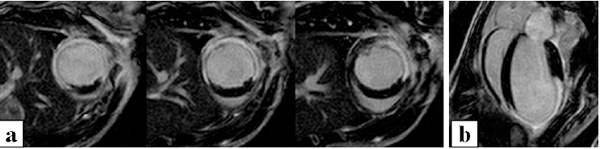
Fig. (8) Permanent occlusion model of myocardial ischaemia in the mouse. Late gadolinium images shown here are acquired 48hrs after myocardial infarction (MI) surgery with 3 consecutive short-axis slices (a), and a 4-chamber long-axis view (b), revealing myocardial ischaemic region and the early stages of wall thinning (figures from reference [61]).