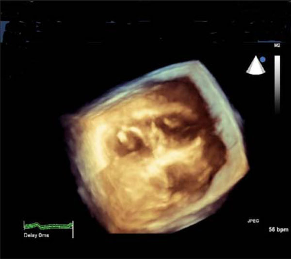
Fig. (2b)
3D-echocardiographic cross-sectional image of the heart: thickened mitral valve (left-side) and papillary muscles of the tricuspid valve (right-side).