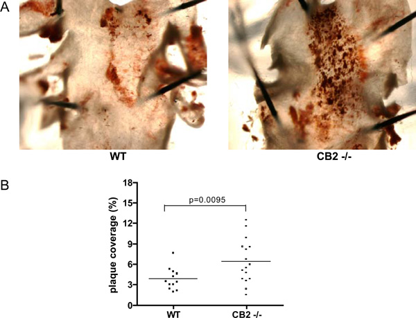
Fig. (2) A) Representative photomicrographs of Oil Red O stained aortic arches derived from a WT transplanted animal (left panel) and a CB2-/- transplanted animal (right panel). Dark red staining indicates an atherosclerotic lesion. B) The atherosclerotic lesion area measured in the aortic arch by quantification of Oil red O staining. Shown is the percentage of the aortic arch covered with an atherosclerotic lesion for each individual animal.