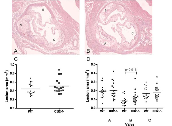
Fig. (3) Representative photomicrographs of the aortic valve area of a WT transplanted LDLr-/- mouse (panel A) and a CB2-/- transplanted LDLr-/- mouse (panel B) stained with haematoxylin-eosin (magnification 20x). Note that in the CB2-/- transplanted animal all three aortic valves are filled with an atherosclerotic plaque, whereas in the WT transplanted animal one aortic valve is virtually free of atherosclerotic lesions. C) The atherosclerotic lesion area measured in the entire aortic valve area. Shown is the average atherosclerotic lesion size in the aortic root for each individual animal. D) The atherosclerotic lesion area measured per aortic valve. Shown is the average atherosclerotic lesion size per aortic valve for each individual animal. The valves were indicated A, B, and C arbitrarily, however, care was taken that for each animal A, B and C had the same orientation in the heart.